
Positive flutter waves in leads II, III, aVF This uncommon variant produces the opposite pattern: Positive flutter waves in V1 – may resemble upright P wavesĬlockwise Reentry. Inverted flutter waves in leads II,III, aVF In this situation the QRS complex is often wide, and the ECG can resemble ventricular tachycardia or ventricular fibrillation.Īnticlockwise Reentry: Commonest form of atrial flutter (90% of cases). Ventricular response rates as high as 180 bpm can occur in patients with normal AV node function.Įxtremely rapid ventricular responses in excess of 180 bpm can be seen in patients with accessory AV nodal bypass tracts. In many instances, treatment of the underlying disease process restores sinus rhythm.

It occurs in approximately 30% of patients with AF and may be associated with more intense symptoms than AF because of the more rapid ventricular response.Ībout 60% of patients experience atrial flutter in association with an acute exacerbation of a chronic condition such as Characteristically the ventricular rate is about 150 bpm.Ītrial flutter frequently occurs in association with other dysrhythmias such as AF. Most commonly, patients have 2:1 AV conduction an atrial rate of 300 bpm with 2:1 conduction, for example, results in a ventricular rate of 150 bpm.

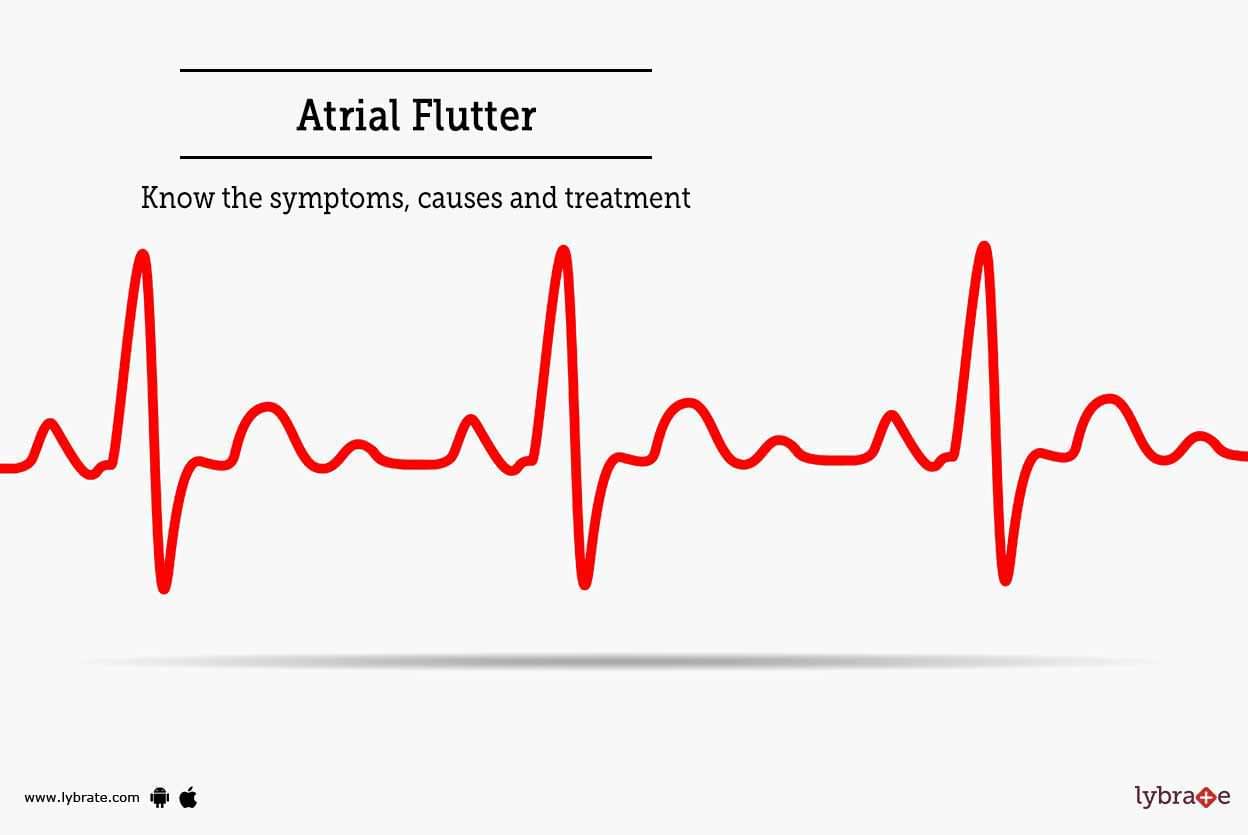
The ventricular rate may be regular or irregular depending on the rate of conduction. The flutter waves are not separated by an isoelectric baseline. The rapid P waves create a sawtooth appearance on ECG and are called flutter waves.įlutter waves are particularly noticeable in leads II, III, aVF, and V1. The length of the re-entry circuit corresponds to the size of the right atrium, resulting in a fairly predictable atrial rate of around 300 bpm (range 200-400). An organized atrial rhythm with an atrial rate of 250–350 bpm with varying degrees of AV block.Ītrial flutter is a type of supraventricular tachycardia caused by a re-entry circuit within the right atrium.


 0 kommentar(er)
